Overview
Bronchogenic cysts are congenital in nature. They are part of a spectrum of congenital abnormalities of the lung, including pulmonary sequestration, congenital cystic adenomatoid malformation, and congenital lobar hyperinflation (emphysema) (see Images below).[1, 2, 3]
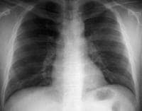 Bronchogenic cyst. Anteroposterior view on conventional radiograph demonstrates a mass in the aorto-pulmonary window.
 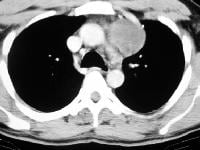 Bronchogenic cyst. Anteroposterior CT demonstrates a mass with fluid density.
 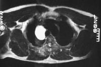 Bronchogenic cyst. Axial T2-weighted MRI demonstrates a high signal mass in the right paratracheal region.
CT scan findings are characteristic when the lesion demonstrates water density. If the lesion demonstrates soft-tissue density, differentiating the cyst from lymph nodes or other solid lesions is difficult.
MRI findings are usually diagnostic for mediastinal cysts.
Intrapulmonary cysts are difficult to diagnose and must usually be aspirated to confirm the diagnosis.
Bronchogenic cyst. Anteroposterior view on conventional radiograph demonstrates a mass in the aorto-pulmonary window. Bronchogenic cyst. Anteroposterior CT demonstrates a mass with fluid density. Bronchogenic cyst. Axial T2-weighted MRI demonstrates a high signal mass in the right paratracheal region. Preferred examination
Bronchogenic cysts are usually an incidental finding, and differentiating them from other pathologic conditions is important. On conventional radiographs, the appearances of mediastinal or lung masses are nonspecific and should be evaluated further using computed tomography (CT) scanning or magnetic resonance imaging (MRI).[4, 5, 6]Limitations of techniques
Chest radiography is usually adequate for detecting larger mediastinal or lung masses; however, it is limited in its ability to differentiate solid masses from fluid.CT scan findings are characteristic when the lesion demonstrates water density. If the lesion demonstrates soft-tissue density, differentiating the cyst from lymph nodes or other solid lesions is difficult.
MRI findings are usually diagnostic for mediastinal cysts.
Intrapulmonary cysts are difficult to diagnose and must usually be aspirated to confirm the diagnosis.
Radiography
Mediastinal cysts are visualized as a mediastinal mass on conventional radiographs.[4] Intrapulmonary cysts usually present as a solitary pulmonary nodule unless the cyst contains air. Radiographs of bronchogenic cysts are depicted in the images below.
Bronchogenic cyst. Anteroposterior view on conventional radiograph demonstrates a mass in the aorto-pulmonary window. 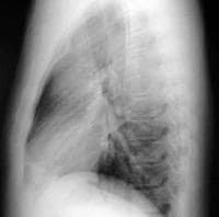 Bronchogenic cyst. Lateral view on conventional radiograph demonstrates filling of the retrosternal clear space correlating to the abnormality observed on the frontal view.
 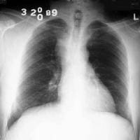 Bronchogenic cyst. Conventional radiograph demonstrates a right paratracheal mass.
 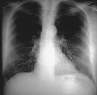 Bronchogenic cyst. Conventional radiograph demonstrates a subcarinal mass.
 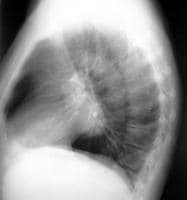 Bronchogenic cyst. Conventional radiograph demonstrates a subcarinal mass.
 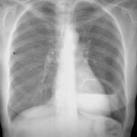 Bronchogenic cyst. Conventional radiograph demonstrates a thin-walled cyst in the left lower lobe with an air-fluid level.
Bronchogenic cyst. Anteroposterior view on conventional radiograph demonstrates a mass in the aorto-pulmonary window. Bronchogenic cyst. Lateral view on conventional radiograph demonstrates filling of the retrosternal clear space correlating to the abnormality observed on the frontal view. Bronchogenic cyst. Conventional radiograph demonstrates a right paratracheal mass. Bronchogenic cyst. Conventional radiograph demonstrates a subcarinal mass. Bronchogenic cyst. Conventional radiograph demonstrates a subcarinal mass. Bronchogenic cyst. Conventional radiograph demonstrates a thin-walled cyst in the left lower lobe with an air-fluid level. Degree of confidence
On conventional radiographs, findings are nonspecific. Mediastinal masses should be evaluated further using CT scanning or MRI to confirm the presence of fluid.False positives/negatives
Difficulty is encountered in determining whether the visualized mass is benign (eg, a bronchogenic cyst) or malignant.Computed Tomography
Bronchogenic cysts are sharply marginated masses demonstrating water or soft-tissue density.[4, 6] Differences in attenuation result from the amount of proteinaceous fluid within the cysts. Cysts do not enhance after administration of IV contrast. An article from the Armed Forces Institute of Pathology documented the appearance of 62 cysts: 40% were water density, 40% were soft-tissue density, 5% contained milk of calcium, 10% were indeterminate from streak artifact, and the remainder were intrapulmonary, either completely air filled or containing an air-fluid level.[7]
In addition to intrapulmonary and mediastinal locations, bronchogenic cysts have been reported to be located in infradiaphragmatic areas, cutaneous areas, intrapericardial areas, and intramural areas of the esophagus. CT scans of bronchogenic cysts are depicted in the images below.
Bronchogenic cyst. Anteroposterior CT demonstrates a mass with fluid density. 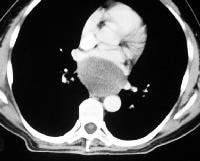 Bronchogenic cyst. CT demonstrates a subcarinal mass with fluid density.
 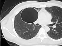 Bronchogenic cyst. CT demonstrates a thin-walled cyst in the right upper lobe.
In addition to intrapulmonary and mediastinal locations, bronchogenic cysts have been reported to be located in infradiaphragmatic areas, cutaneous areas, intrapericardial areas, and intramural areas of the esophagus. CT scans of bronchogenic cysts are depicted in the images below.
Bronchogenic cyst. Anteroposterior CT demonstrates a mass with fluid density. Bronchogenic cyst. CT demonstrates a subcarinal mass with fluid density. Bronchogenic cyst. CT demonstrates a thin-walled cyst in the right upper lobe. Degree of confidence
In the proper clinical setting, a CT scan finding of a sharply marginated, nonenhancing, water-density mass is diagnostic of a bronchogenic cyst. Nonenhancing masses demonstrating soft-tissue density need further evaluation using MRI. Location is also important. Intrapulmonary cysts are usually difficult to diagnose and usually require aspiration for diagnosis.False positives/negatives
Most bronchogenic cysts are relatively characteristic in appearance on CT scan, but in atypical cases with hemorrhage or infection, findings may be confused with those of necrotic adenopathy, cystic lung disease, or lung abscess.Magnetic Resonance Imaging
Bronchogenic cysts are usually bright on T2-weighted images and dark on T1-weighted images (see the Image below).[6] Cysts do not enhance after administration of IV gadolinium.
Bronchogenic cyst. Axial T2-weighted MRI demonstrates a high signal mass in the right paratracheal region.
Bronchogenic cyst. Axial T2-weighted MRI demonstrates a high signal mass in the right paratracheal region.
No comments:
Post a Comment