MRI Technique
In
magnetic resonance imaging (MRI) of the elbow, patients are imaged in
the supine position or in the prone position with the arm overhead.
Imaging begins about 10 cm above the elbow joint and extends to the
bicipital tuberosity. Images are acquired in the axial, coronal, and
sagittal planes.[1, 2]
The authors routinely perform axial and coronal T1-weighted (T1W) spin-echo (SE) imaging; axial, coronal, and sagittal fat-suppressed T2-weighted (T2W) SE or short-tau inversion recovery (STIR) imaging (see the images below); and coronal proton density–weighted imaging by 4-mm section thickness with a 0.4-mm intersection gap.
 Axial
short-tau inversion recovery (STIR) images show hyperintense fluid
around the biceps tendon in a patient with a partial tear of the biceps
tendon.
Axial
short-tau inversion recovery (STIR) images show hyperintense fluid
around the biceps tendon in a patient with a partial tear of the biceps
tendon.  Coronal short-tau inversion recovery (STIR) images demonstrate a partial tear of the radial collateral ligament.
 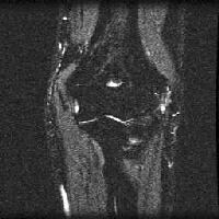 Lateral
epicondylitis. Coronal short-tau inversion recovery (STIR) image
demonstrates edema near the origin of the common extensor. Intravenous
gadolinium-based contrast agent is given to patients with a suspected
elbow mass lesion or infection. About 10 mL of a 1:100-250 mixture of
contrast agent and saline is injected into the joint if magnetic
resonance (MR) arthrography is performed.[3]
Lateral
epicondylitis. Coronal short-tau inversion recovery (STIR) image
demonstrates edema near the origin of the common extensor. Intravenous
gadolinium-based contrast agent is given to patients with a suspected
elbow mass lesion or infection. About 10 mL of a 1:100-250 mixture of
contrast agent and saline is injected into the joint if magnetic
resonance (MR) arthrography is performed.[3]
Gadolinium-based contrast agents have been linked to the development of nephrogenic systemic fibrosis (NSF) or nephrogenic fibrosing dermopathy (NFD). The disease has occurred in patients with moderate to end-stage renal disease after being given a gadolinium-based contrast agent to enhance MRI or MRA scans. NSF/NFD is a debilitating and sometimes fatal disease. Characteristics include red or dark patches on the skin; burning, itching, swelling, hardening, and tightening of the skin; yellow spots on the whites of the eyes; joint stiffness with trouble moving or straightening the arms, hands, legs, or feet; pain deep in the hip bones or ribs; and muscle weakness.
The authors routinely perform axial and coronal T1-weighted (T1W) spin-echo (SE) imaging; axial, coronal, and sagittal fat-suppressed T2-weighted (T2W) SE or short-tau inversion recovery (STIR) imaging (see the images below); and coronal proton density–weighted imaging by 4-mm section thickness with a 0.4-mm intersection gap.
Axial
short-tau inversion recovery (STIR) images show hyperintense fluid
around the biceps tendon in a patient with a partial tear of the biceps
tendon. Coronal short-tau inversion recovery (STIR) images demonstrate a partial tear of the radial collateral ligament. Lateral
epicondylitis. Coronal short-tau inversion recovery (STIR) image
demonstrates edema near the origin of the common extensor. Intravenous
gadolinium-based contrast agent is given to patients with a suspected
elbow mass lesion or infection. About 10 mL of a 1:100-250 mixture of
contrast agent and saline is injected into the joint if magnetic
resonance (MR) arthrography is performed.[3] Gadolinium-based contrast agents have been linked to the development of nephrogenic systemic fibrosis (NSF) or nephrogenic fibrosing dermopathy (NFD). The disease has occurred in patients with moderate to end-stage renal disease after being given a gadolinium-based contrast agent to enhance MRI or MRA scans. NSF/NFD is a debilitating and sometimes fatal disease. Characteristics include red or dark patches on the skin; burning, itching, swelling, hardening, and tightening of the skin; yellow spots on the whites of the eyes; joint stiffness with trouble moving or straightening the arms, hands, legs, or feet; pain deep in the hip bones or ribs; and muscle weakness.
Gross Anatomy
Formed by the distal humerus, proximal ulna, and proximal radius,
the elbow is a hinge-type synovial joint that provides both stability
and function. The distal aspect of the humerus is a wide, flattened
structure. The medial third of its articular surface, the trochlea,
articulates with the ulna, whereas its lateral capitellum
articulates with the radius. A hollow area on the posterior surface of
the humerus, above the trochlea, is termed the olecranon fossa. The
posterior capsular attachment of the humerus is located above this
fossa. (See the images below.)[4, 5, 6]
 Coronal short-tau inversion recovery (STIR) image demonstrates a normal ulnar collateral ligament.
 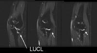 Coronal
short-tau inversion recovery (STIR) image shows a normal radial
collateral ligament. LUCL = lateral ulnar collateral ligament, a part of
radial collateral ligament. The anterior aspect of the distal humerus contains 2 fossae: the coronoid
fossa, located medially, and the radial fossa, located laterally. The
anterior capsular attachment to the humerus is located above these
fossae.
Coronal
short-tau inversion recovery (STIR) image shows a normal radial
collateral ligament. LUCL = lateral ulnar collateral ligament, a part of
radial collateral ligament. The anterior aspect of the distal humerus contains 2 fossae: the coronoid
fossa, located medially, and the radial fossa, located laterally. The
anterior capsular attachment to the humerus is located above these
fossae.
The proximal end of the ulna contains 2 processes: the olecranon and the coronoid. The olecranon process is posteriorly smooth at the site of attachment of the triceps tendon. The proximal end of the radius consists of a head, a neck, and a tuberosity. The radial head is disk shaped and contains a shallow, cupped articular surface that articulates with the capitellum of the humerus. The radial tuberosity is located beneath the medial aspect of the neck.
A thin, broad, and weak fibrous capsule envelops the entire elbow. The capsule anteriorly extends from its attachment sites above the distal humeral fossae to its distal attachment to the coronoid process and annular ligament. In its posterior portion, the capsule attaches to the capitellum, the olecranon fossa, and the medial epicondyle. At its inferomedial aspect, the capsule attaches to the upper and lateral margins of the olecranon. The lateral part of the capsule is continuous with the superior radioulnar joint. The overlying collateral ligaments strengthen the capsule.
The synovial membrane of the elbow lines the deep surface of the fibrous capsule and the annular ligament. It extends from the articular surface of the humerus, contacting with the olecranon, distal humeral fossae, and medial trochlea surface. A synovial fold projects into the joint between the radius and ulna, partially dividing the articulation into humeroulnar and humeroradial portions.
Several fat pads are located between the fibrous capsule and the synovial membrane. Fat pads are near the synovial fold between the radius and ulna and over the olecranon, coronoid, and radial fossae. These fat pads are extrasynovial but intracapsular. The anterior fat pad, anterior to the distal humerus, normally assumes a teardrop configuration. On lateral elbow radiographs, with the elbow at 90° of flexion, this anterior fat pad is visible. On the contrary, the posterior fat pad, in the olecranon fossa, is normally not visible on lateral radiographs with the elbow in 90° of flexion. However, intra-articular processes that expand the elbow joint (eg, joint effusion) displace these pads.
Radial collateral ligaments and ulnar collateral ligaments (UCLs) reinforce the fibrous capsule.
The radial or lateral collateral ligamentous (LCL) complex consists of the radial collateral ligament, annular ligament, lateral UCL (LUCL), and accessory collateral ligament. The radial collateral ligament attaches superiorly to the lateral epicondyle and inferiorly to the radial notch of the ulna and to the annular ligament.
The ulnar or medial collateral ligament (MCL) is composed of 3 distinct yet continuous bands. The anterior band spans the distance between the medial epicondyle and the coronoid process. The posterior band arises from the posterior aspect of the medial epicondyle to the medial edge of the olecranon process. A thin intermediate band merges with the adjacent bands by virtue of an oblique ray of fibers.
The superior radioulnar joint is located between the radial head and a fibro-osseous ring formed by the annular ligament and the radial notch of the ulna. The radioulnar joint is lined with articular cartilage that is contiguous with the trochlear notch. The radial head is also lined with articular cartilage. The annular ligament anteriorly attaches to the anterior margin of the radial notch. It encircles the head of the radius. At the posterior aspect, it contains several bands that attach to the ulna near the posterior margin of the radial notch.
Coronal short-tau inversion recovery (STIR) image demonstrates a normal ulnar collateral ligament. Coronal
short-tau inversion recovery (STIR) image shows a normal radial
collateral ligament. LUCL = lateral ulnar collateral ligament, a part of
radial collateral ligament. The anterior aspect of the distal humerus contains 2 fossae: the coronoid
fossa, located medially, and the radial fossa, located laterally. The
anterior capsular attachment to the humerus is located above these
fossae. The proximal end of the ulna contains 2 processes: the olecranon and the coronoid. The olecranon process is posteriorly smooth at the site of attachment of the triceps tendon. The proximal end of the radius consists of a head, a neck, and a tuberosity. The radial head is disk shaped and contains a shallow, cupped articular surface that articulates with the capitellum of the humerus. The radial tuberosity is located beneath the medial aspect of the neck.
A thin, broad, and weak fibrous capsule envelops the entire elbow. The capsule anteriorly extends from its attachment sites above the distal humeral fossae to its distal attachment to the coronoid process and annular ligament. In its posterior portion, the capsule attaches to the capitellum, the olecranon fossa, and the medial epicondyle. At its inferomedial aspect, the capsule attaches to the upper and lateral margins of the olecranon. The lateral part of the capsule is continuous with the superior radioulnar joint. The overlying collateral ligaments strengthen the capsule.
The synovial membrane of the elbow lines the deep surface of the fibrous capsule and the annular ligament. It extends from the articular surface of the humerus, contacting with the olecranon, distal humeral fossae, and medial trochlea surface. A synovial fold projects into the joint between the radius and ulna, partially dividing the articulation into humeroulnar and humeroradial portions.
Several fat pads are located between the fibrous capsule and the synovial membrane. Fat pads are near the synovial fold between the radius and ulna and over the olecranon, coronoid, and radial fossae. These fat pads are extrasynovial but intracapsular. The anterior fat pad, anterior to the distal humerus, normally assumes a teardrop configuration. On lateral elbow radiographs, with the elbow at 90° of flexion, this anterior fat pad is visible. On the contrary, the posterior fat pad, in the olecranon fossa, is normally not visible on lateral radiographs with the elbow in 90° of flexion. However, intra-articular processes that expand the elbow joint (eg, joint effusion) displace these pads.
Radial collateral ligaments and ulnar collateral ligaments (UCLs) reinforce the fibrous capsule.
The radial or lateral collateral ligamentous (LCL) complex consists of the radial collateral ligament, annular ligament, lateral UCL (LUCL), and accessory collateral ligament. The radial collateral ligament attaches superiorly to the lateral epicondyle and inferiorly to the radial notch of the ulna and to the annular ligament.
The ulnar or medial collateral ligament (MCL) is composed of 3 distinct yet continuous bands. The anterior band spans the distance between the medial epicondyle and the coronoid process. The posterior band arises from the posterior aspect of the medial epicondyle to the medial edge of the olecranon process. A thin intermediate band merges with the adjacent bands by virtue of an oblique ray of fibers.
The superior radioulnar joint is located between the radial head and a fibro-osseous ring formed by the annular ligament and the radial notch of the ulna. The radioulnar joint is lined with articular cartilage that is contiguous with the trochlear notch. The radial head is also lined with articular cartilage. The annular ligament anteriorly attaches to the anterior margin of the radial notch. It encircles the head of the radius. At the posterior aspect, it contains several bands that attach to the ulna near the posterior margin of the radial notch.
Tendinous, Ligamentous, and Muscle Abnormalities
Soft-tissue
abnormalities involving the tendons, ligaments, and muscles include
rupture of the biceps and/or triceps tendons, lateral or medial
epicondylitis, injury to the lateral collateral ligament (LCL) or medial
collateral ligament (MCL), and posterior dislocation of elbow.
Disruptions of the ligaments about the elbow can accompany severe physical trauma (eg, elbow dislocation),[7] less-extensive acute trauma (eg, valgus injury),[8] or chronic stress (eg, pitching a baseball). Injuries to the UCL or MCL predominate.
The ulnar collateral ligament (UCL) or MCL is the primary structure that maintains elbow stability in the presence of valgus stress, though other structures, such as the adjacent flexor musculature, joint capsule, and articular surfaces, contribute to stability. The anterior band of the UCL is functionally the most important for the elbow joint.
On T1W MRIs, findings include increased signal intensity suggesting heterotopic bone formation near the medial epicondyle, epicondylar avulsion, formation of traction osteophytes or subchondral cysts, or sclerosis in capitellum due to lateral injury. On T2W images, findings include increased signal intensity in the anterior band of the MCL, hyperintensity or hypertrophy of the ulnar insertion site (called a sublime tubercle) with or without a full-thickness tear and retraction.
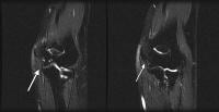
Images of a chronically injured UCL demonstrate laxity, irregularity, and poor definition without increased signal intensity around the ligament. Midsubstance ruptures are most common. Partial detachment of deep undersurface of anterior bundle of UCL should be evaluated by using MR arthrograms, which demonstrate contrast enhancement extending around the corner of sublime tubercle. Images of complete tears show frank extravasation of contrast agent. The differential diagnosis includes medial epicondylitis and a sublime tubercle fracture. (See the image below.)
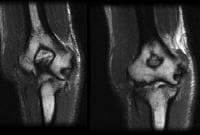 Coronal T1-weighted (T1W) images show a partial tear of the ulnar collateral ligament. LCL
injury is less common than MCL injury. LCL injury is usually the result
of chronic trauma, and it is associated with tennis elbow. LUCL injury
is most important, as it results in posterolateral rotatory
insufficiency. Laxity can occur after extensive subperiosteal elevation
of the extensor tendons in cases of tennis elbow. On T1W images, a tear
of the LUCL ligament may be seen at its humeral origin, and on T2W
images, disruption of ligamentous fibers with fluid signal intensity may
be seen. Associated findings include fractures of coronoid process,
radial head, or capitellum and dislocation of the radius or ulna.
Coronal T1-weighted (T1W) images show a partial tear of the ulnar collateral ligament. LCL
injury is less common than MCL injury. LCL injury is usually the result
of chronic trauma, and it is associated with tennis elbow. LUCL injury
is most important, as it results in posterolateral rotatory
insufficiency. Laxity can occur after extensive subperiosteal elevation
of the extensor tendons in cases of tennis elbow. On T1W images, a tear
of the LUCL ligament may be seen at its humeral origin, and on T2W
images, disruption of ligamentous fibers with fluid signal intensity may
be seen. Associated findings include fractures of coronoid process,
radial head, or capitellum and dislocation of the radius or ulna.
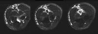
Discontinuity and retraction of the tendon accompany complete tears, whereas remaining intact tendinous fibers accompany partial tears. Alterations in signal intensity depend on the age of the injury. On proton density–weighted images, tendinosis is seen as increased signal intensity in a thickened tendon. On T2W MRIs, increased signal intensity similar to that of fluid without discontinuity of the tendon suggests partial tear. Full-thickness tears with or without retraction of the ends of the tendon indicate a complete tear. Sagittal fat-suppressed T2W images are particularly useful in identifying tendon retraction in injuries of the biceps and triceps tendons. (See the images below.)
Axial
short-tau inversion recovery (STIR) images show hyperintense fluid
around the biceps tendon in a patient with a partial tear of the biceps
tendon. 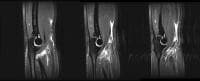 Sagittal short-tau inversion recovery (STIR) images demonstrate a complete tear and avulsion of the biceps tendon. Although
rupture of the distal portion of the tendon of the biceps brachii
muscle accounts for only 5% of all biceps injuries, they are an
important injury about the elbow. The typical mechanism is related to
forceful hyperextension applied to a flexed and supinated forearm.
Complete rupture, with avulsion of the tendinous attachment to the
radial tuberosity, generally occurs in men older than 40 years and in
the dominant extremity.
Sagittal short-tau inversion recovery (STIR) images demonstrate a complete tear and avulsion of the biceps tendon. Although
rupture of the distal portion of the tendon of the biceps brachii
muscle accounts for only 5% of all biceps injuries, they are an
important injury about the elbow. The typical mechanism is related to
forceful hyperextension applied to a flexed and supinated forearm.
Complete rupture, with avulsion of the tendinous attachment to the
radial tuberosity, generally occurs in men older than 40 years and in
the dominant extremity.
Associated findings are a fluid-filled bicipital bursa and tendon sheath, a hypointense and thickened tendon on T1W images, and hypertrophy of the radial tuberosity. Lateral epicondylitis is sometimes present.[9] An important differential diagnosis is bicipital radial bursitis, which results in fluid in the bicipital bursa with normal signal intensity in the adjacent tendon. An absence of signal-intensity changes on T2W and STIR images with an atrophic or hypertrophic tendon indicate a chronic tear.
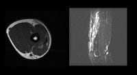 Axial T1- and sagittal T2-weighted fat-saturated images show a sprain of the triceps muscle and tendinosis.
Coronal short-tau inversion recovery (STIR) images demonstrate a partial tear of the radial collateral ligament. Lateral
epicondylitis. Coronal short-tau inversion recovery (STIR) image
demonstrates edema near the origin of the common extensor.
Disruptions of the ligaments about the elbow can accompany severe physical trauma (eg, elbow dislocation),[7] less-extensive acute trauma (eg, valgus injury),[8] or chronic stress (eg, pitching a baseball). Injuries to the UCL or MCL predominate.
The ulnar collateral ligament (UCL) or MCL is the primary structure that maintains elbow stability in the presence of valgus stress, though other structures, such as the adjacent flexor musculature, joint capsule, and articular surfaces, contribute to stability. The anterior band of the UCL is functionally the most important for the elbow joint.
On T1W MRIs, findings include increased signal intensity suggesting heterotopic bone formation near the medial epicondyle, epicondylar avulsion, formation of traction osteophytes or subchondral cysts, or sclerosis in capitellum due to lateral injury. On T2W images, findings include increased signal intensity in the anterior band of the MCL, hyperintensity or hypertrophy of the ulnar insertion site (called a sublime tubercle) with or without a full-thickness tear and retraction.
Images of a chronically injured UCL demonstrate laxity, irregularity, and poor definition without increased signal intensity around the ligament. Midsubstance ruptures are most common. Partial detachment of deep undersurface of anterior bundle of UCL should be evaluated by using MR arthrograms, which demonstrate contrast enhancement extending around the corner of sublime tubercle. Images of complete tears show frank extravasation of contrast agent. The differential diagnosis includes medial epicondylitis and a sublime tubercle fracture. (See the image below.)
Coronal T1-weighted (T1W) images show a partial tear of the ulnar collateral ligament. LCL
injury is less common than MCL injury. LCL injury is usually the result
of chronic trauma, and it is associated with tennis elbow. LUCL injury
is most important, as it results in posterolateral rotatory
insufficiency. Laxity can occur after extensive subperiosteal elevation
of the extensor tendons in cases of tennis elbow. On T1W images, a tear
of the LUCL ligament may be seen at its humeral origin, and on T2W
images, disruption of ligamentous fibers with fluid signal intensity may
be seen. Associated findings include fractures of coronoid process,
radial head, or capitellum and dislocation of the radius or ulna. Avulsions and tears
Avulsions and tears of tendons about the elbow are uncommon. Avulsion at sites of tendinous attachment may occur as a complication of systemic diseases, such as primary and secondary hyperparathyroidism, systemic lupus erythematosus, rheumatoid arthritis, and osteogenesis imperfecta. They may also occur as a consequence of physical injury. Tears of the tendon can be graded as follows: grade I is mild strain and/or tendinosis, grade II is partial tear, and grade III is complete disruption.Discontinuity and retraction of the tendon accompany complete tears, whereas remaining intact tendinous fibers accompany partial tears. Alterations in signal intensity depend on the age of the injury. On proton density–weighted images, tendinosis is seen as increased signal intensity in a thickened tendon. On T2W MRIs, increased signal intensity similar to that of fluid without discontinuity of the tendon suggests partial tear. Full-thickness tears with or without retraction of the ends of the tendon indicate a complete tear. Sagittal fat-suppressed T2W images are particularly useful in identifying tendon retraction in injuries of the biceps and triceps tendons. (See the images below.)
Axial
short-tau inversion recovery (STIR) images show hyperintense fluid
around the biceps tendon in a patient with a partial tear of the biceps
tendon. Sagittal short-tau inversion recovery (STIR) images demonstrate a complete tear and avulsion of the biceps tendon. Although
rupture of the distal portion of the tendon of the biceps brachii
muscle accounts for only 5% of all biceps injuries, they are an
important injury about the elbow. The typical mechanism is related to
forceful hyperextension applied to a flexed and supinated forearm.
Complete rupture, with avulsion of the tendinous attachment to the
radial tuberosity, generally occurs in men older than 40 years and in
the dominant extremity. Associated findings are a fluid-filled bicipital bursa and tendon sheath, a hypointense and thickened tendon on T1W images, and hypertrophy of the radial tuberosity. Lateral epicondylitis is sometimes present.[9] An important differential diagnosis is bicipital radial bursitis, which results in fluid in the bicipital bursa with normal signal intensity in the adjacent tendon. An absence of signal-intensity changes on T2W and STIR images with an atrophic or hypertrophic tendon indicate a chronic tear.
Triceps tendon injuries
Regarding injuries of the triceps tendon, the typical patient is a middle-aged man who had forced flexion of an extended forearm as in a motorcycle accident or sports-related trauma. Most tears occur at the insertion site at the olecranon process.[10] Avulsion of the tendo-osseous attachment may be observed. Myotendinous ruptures are uncommon. Other associated abnormalities are olecranon bursitis and radial-head fractures. The differential diagnosis includes olecranon bursitis, which may be associated with infection and in which the underlying tendon appears normal; hematoma of the triceps muscle, where the muscle appears hyperintense on T1W and T2W images; and fracture of the olecranon, which commonly occur in adolescent due to direct trauma or a throwing injury. (See the image below.)Axial T1- and sagittal T2-weighted fat-saturated images show a sprain of the triceps muscle and tendinosis. Medial epicondylitis
Medial epicondylitis is also known as golfer's elbow or medial tennis elbow. This injury is due to chronic overload and overuse of flexor pronator muscle group. It involves the interface between the origins of the pronator teres and the flexor carpi radialis. Coronal fat-suppressed T2W images demonstrate hyperintensity of the origin of flexor pronator.Lateral epicondylitis
Lateral epicondylitis, or tennis elbow, is 7-20 times more common than medial epicondylitis. This injury is characterized by degeneration and partial tears of the extensor group of tendons. Partial avulsion of the radial collateral ligament and the posterior elbow dislocation may also be present. Hyperintensity is seen on fat-suppressed T2W or STIR images in the origin of the common extensor. Associated periostitis, increased signal intensity in the anconeus muscle, and soft-tissue edema may be seen. (See the images below.)Coronal short-tau inversion recovery (STIR) images demonstrate a partial tear of the radial collateral ligament. Lateral
epicondylitis. Coronal short-tau inversion recovery (STIR) image
demonstrates edema near the origin of the common extensor. Neuropathies Around The Elbow Joint
Entrapment and compression neuropathies about the elbow may involve the ulnar, radial, or median nerve.
MRI abnormalities include displacement or shift of the ulnar nerve, a soft tissue mass, and thickening and increased signal intensity in the compressed nerve. Ulnar-nerve enlargement may be focal, occurring at the level of the cubital tunnel, with normal girth noted proximal and distal to the tunnel. On fat-suppressed T2W images, neurogenic edema can be seen in muscles the ulnar nerve supplies. A thickened cubital tunnel retinaculum may be demonstrated. The differential diagnosis includes enlarged perineural veins, which due to increased venous pressure are seen as flow voids on T2W images.
In cases of compression of the radial or median nerve, MRI may reveal a mass (eg, lipoma, ganglion cyst) and displacement and abnormal signal intensity in the affected nerve.
Ulnar nerve
Neuropathies involving the ulnar nerve are the most common. Entrapment of the ulnar nerve in the fibro-osseous tunnel posterior to the medial epicondyle of the humerus results in the cubital tunnel syndrome. Typical causes of this syndrome are injury and progressive cubitus valgus deformity (tardy ulnar nerve palsy). Other causes are osteoarthritis, rheumatoid arthritis, nerve subluxation, prolonged bedrest, anomalous muscles, thickened cubital tunnel retinaculum (Osborne ligament), malunion or nonunion of a condylar fracture, masses, and trochlear hypoplasia.MRI abnormalities include displacement or shift of the ulnar nerve, a soft tissue mass, and thickening and increased signal intensity in the compressed nerve. Ulnar-nerve enlargement may be focal, occurring at the level of the cubital tunnel, with normal girth noted proximal and distal to the tunnel. On fat-suppressed T2W images, neurogenic edema can be seen in muscles the ulnar nerve supplies. A thickened cubital tunnel retinaculum may be demonstrated. The differential diagnosis includes enlarged perineural veins, which due to increased venous pressure are seen as flow voids on T2W images.
Radial nerve
Entrapment of a portion of the radial nerve may occur just distal to the elbow, where the posterior interosseous, or deep, branch passes into the supinator muscle (arcade of Frohse). Individuals in occupations that require frequent supination and pronation are susceptible to this injury. Causes of supinator syndrome are elbow dislocations, fractures, rheumatoid arthritis, soft tissue tumors, and traumatic or developmental fibrous bands. On fat-suppressed T2W images, findings include diffuse increased signal intensity in all or some muscles the posterior interosseus nerve supplies; this finding suggests denervation. Reduced size of the muscle belly and fatty atrophy may be seen. The differential diagnosis includes nonspecific myositis; T2W images may show hyperintensity in the muscle, which is related to trauma.Median nerve
Median-nerve entrapment can occur due to hypertrophy of the pronator teres muscle or fibrous bands leading to compression of the nerve in the antecubital area where the nerve passes between the 2 heads of the pronator teres muscle under the edge of the flexor digitorum sublimis muscle (pronator syndrome). This syndrome results in sensory symptoms as opposed to anterior interosseus nerve (AIN) syndrome, which involves denervation of flexor pollicis longus and flexor digitorum profundus to second and third digits. T2W and STIR images demonstrate diffuse increase signal intensity in affected muscles, with decreased muscle bulk suggesting denervation. Fatty atrophy is best demonstrated on T1W images.In cases of compression of the radial or median nerve, MRI may reveal a mass (eg, lipoma, ganglion cyst) and displacement and abnormal signal intensity in the affected nerve.
Bone Abnormalities
Osteochondritis dissecans
Osteochondritis dissecans about the elbow usually affects the capitulum in boys and male adolescents aged 12-16 years. This disorder differs from a developmental alteration (osteochondrosis) of the capitellar ossification center known as Panner disease, which affects children aged 5-11 years. The latter disease process affects the whole capitellum, and loose body formation and residual deformity is usually absent in Panner disease. (See the image below.)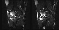 Coronal
short-tau inversion recovery (STIR) images demonstrate osteochondritis
dissecans with adjoining marrow edema in the capitulum. The
precise relationship of osteochondritis dissecans of the capitulum and
an osteochondral fracture is not clear, but most investigators regard
the former as a posttraumatic abnormality that may lead to
osteonecrosis. MRI of osteochondritis dissecans of the capitulum may be
performed to gain information about the integrity of the adjacent
articular cartilage, the viability of the separated fragment, and the
presence of associated intra-articular osseous and cartilaginous bodies.
Variable signal intensity may be seen in the fragment depending on the
degree of sclerosis. Cystic changes may be seen. T1W images may show
increased signal intensity in the fragment, as opposed to normal marrow
fat intensity and hypointensity around the fragment. Joint fluid or
granulation tissue at the interface between the fragment and the parent
bone manifests as increased signal intensity on T2W MRIs and generally
indicates an unstable lesion.
Coronal
short-tau inversion recovery (STIR) images demonstrate osteochondritis
dissecans with adjoining marrow edema in the capitulum. The
precise relationship of osteochondritis dissecans of the capitulum and
an osteochondral fracture is not clear, but most investigators regard
the former as a posttraumatic abnormality that may lead to
osteonecrosis. MRI of osteochondritis dissecans of the capitulum may be
performed to gain information about the integrity of the adjacent
articular cartilage, the viability of the separated fragment, and the
presence of associated intra-articular osseous and cartilaginous bodies.
Variable signal intensity may be seen in the fragment depending on the
degree of sclerosis. Cystic changes may be seen. T1W images may show
increased signal intensity in the fragment, as opposed to normal marrow
fat intensity and hypointensity around the fragment. Joint fluid or
granulation tissue at the interface between the fragment and the parent
bone manifests as increased signal intensity on T2W MRIs and generally
indicates an unstable lesion.Iwasaki et al studied 10 young male athletes with advanced lesions of capitellar osteochondritis dissecans to determine whether MRI findings improve with increasing time after mosaicplasty. The authors found that at 12 months, all patients were able to return to their competitive level of sports. Fluid surrounding the graft was found in all patients at 3 months and in 4 patients at 6 months. At 12 months, the grafts were all well seated within the recipient sites, with no MRI evidence of graft loosening. The overall MRI scores significantly improved from 3 to 12 months. The MRI findings therefore indicated that the graft incorporation to the surrounding tissues occurred around or after 6 months after surgery, suggesting that rehabilitation precautions be taken for up to 6 months after mosaicplasty for young athletes with capitellar OCD.[1]
Han et al evaluated the distribution of shoulder and elbow injuries confirmed by MRI in 554 throwing athletes and found that there is significant difference in the distribution of injuries according to the player's age and position. Junior high school players sustained a higher proportion of osteochondritis dissecans than players in high school and college, and the players in junior high school with UCL injuries were taller and heavier than the players in the control group. High school and college players were more likely to have UCL injuries or SLAP lesions, and in the high school group with UCL injuries or SLAP lesions, the players were both taller and heavier than the players in the control group. Pitchers and outfielders were more likely to have UCL injuries than infielders.[11]
A potential pitfall is a pseudodefect of the capitulum, which is seen as abrupt slope of posterior portion of capitellum that is not associated with any marrow edema in the adjacent bone. Osteochondritis dissecans also occurs on the anterior surface.
Occult fractures
Radiographically occult fracture can be imaged by using MRI, which demonstrates bone-marrow edema with or without a hypointense linear fracture line. Occult fractures are most commonly seen in the region of the radial head in adults and in the supracondylar region in children.A fracture of the coronoid process is due to bony avulsion of the brachialis insertion, and posterior dislocation of the ulna and an olecranal fracture due to direct trauma or forced flexion on an extended forearm may be seen.
Fractures of the lateral condyle result from avulsion injury due to the forearm extensor and supinator muscles, fractures involving the medial condyle result from an avulsion injury due to contraction of the forearm flexor muscles.
Osteomyelitis
Osteomyelitis is commonly seen as an acute process in children and as a chronic process in adults. It is often the result of adjacent infectious bursitis. Staphylococcus aureus is the most common infecting organism in all age groups.MRI findings include bone-marrow edema, which appears hypointense on T1W images and hyperintense on T2W images; interosseus abscess, which appears hyperintense on T2W images; cortical destruction; cloaca, which appears as focal hyperintensity in a hypointense periosteum on T2W images; a Brodie abscess, which appears as focal hyperintensity surrounded by a hypointense sclerotic rim; and cellulitis, myositis, bursitis, and involvement of the sinus tracts. Contrast-enhanced T1W images may show enhancing bone marrow and peripheral enhancement in the abscess.
Synovial Abnormalities
Synovial proliferation in the elbow may accompany a variety of disease processes, including rheumatoid arthritis, septic arthritis, crystal deposition disorders, pigmented villonodular synovitis, and idiopathic synovial osteochondromatosis.
Synovial inflammation and fluid accumulation in the olecranon bursa is seen after injury and in cases of rheumatoid arthritis, septic bursitis, and gout and other diseases involving crystal deposition. The diagnosis of olecranon bursitis is generally apparent on physical examination. A sympathetic effusion in the elbow joint may accompany this condition. In rheumatoid arthritis, subcutaneous nodules may simulate the appearance of olecranon bursitis.
Para-articular synovial cysts that communicate with the elbow joint are a recognized manifestation of rheumatoid arthritis. However, theoretically, they could occur with any process leading to elevation of intra-articular pressure in the elbow. A valvelike mechanism may be present between the synovial cyst and the elbow joint. Such cysts predominate in the antecubital region.
Inflammatory disorders
Rheumatoid arthritis and other synovial inflammatory disorders affecting the elbow lead to proliferation and thickening of the synovial membrane and accumulation of joint fluid. The signal-intensity characteristics of the abnormal synovium and fluid are similar on MRIs obtained with standard sequences. Erosions may be seen as hyperintense defects in the subchondral bone at the joint edges (bare areas). The intravenous administration of gadolinium compounds enhances the signal intensity of the inflammatory tissue and does not affect the signal intensity of fluid on MRIs obtained immediately after their injection.Synovial inflammation and fluid accumulation in the olecranon bursa is seen after injury and in cases of rheumatoid arthritis, septic bursitis, and gout and other diseases involving crystal deposition. The diagnosis of olecranon bursitis is generally apparent on physical examination. A sympathetic effusion in the elbow joint may accompany this condition. In rheumatoid arthritis, subcutaneous nodules may simulate the appearance of olecranon bursitis.
Para-articular synovial cysts that communicate with the elbow joint are a recognized manifestation of rheumatoid arthritis. However, theoretically, they could occur with any process leading to elevation of intra-articular pressure in the elbow. A valvelike mechanism may be present between the synovial cyst and the elbow joint. Such cysts predominate in the antecubital region.
Pigmented villonodular synovitis
Pigmented villonodular synovitis may involve the elbow, initially leading to an effusion and subsequently leading to osteoporosis and even joint-space loss and bone erosions. Although an appropriate clinical history and typical radiographic abnormalities may suggest the diagnosis, the deposition of hemosiderin in the affected synovial tissue produces regions of persistent hypointensity on T2W images, especially, gradient-echo MRIs. This finding also may be observed in cases of chronic hemarthrosis, hemophilia, and synovial hemangioma.Idiopathic synovial osteochondromatosis
Idiopathic synovial osteochondromatosis, which is related to metaplasia of the synovial lining, is accompanied by synovial proliferation and the formation of intrasynovial nodules of cartilage. The fate of these nodules varies, though they may become calcified or ossified. They may become free within the joint cavity and later become embedded in a distant synovial site. Routine radiography is sensitive to calcified and ossified intra-articular bodies, but it is insensitive to nonossified bodies. Arthrography, in which the loose bodies appear as multiple filling defects within the opacified joint, and MRI can be helpful in diagnosing this condition.Loose bodies
Intra-articular and osteocartilaginous loose bodies can result from any process leading to disintegration of the articular surface of the elbow joint. Although trauma is the most important cause, other etiologies include osteoarthritis, synovial osteochondromatosis, and calcium pyrophosphate deposition disease (CPPD). In common with intra-articular bodies in other locations, bodies originating from the joint surface may be composed of cartilage alone, cartilage and bone together, or (in rare cases) bone alone.Bursitis and Other Soft Tissue Masses Around Elbow Joint
Bicipitoradial bursitis
and interosseous bursitis can affect the insertion of the biceps
tendon, as well as flexion and supination of forearm. They typically
occur in men after athletic activity and manifest as a painful
antecubital soft-tissue mass with painful flexion. Findings on MRI
include a cystic mass interposed between the distal biceps tendon and
the anterior aspect of the radial tuberosity. Sometimes, communication
with the elbow joint below the annular ligament may be demonstrated.
Olecranal bursitis occurs in an adolescent or adult patient and appears as a fluid collection posterior to the insertion of the triceps tendon.
MRIs may show soft tissue tumors around the elbow. Some of the tumors may have typical patterns of signal intensity: Lipomas may have fat intensity; liposarcomas, variable signal intensity with or without fat intensity; ganglion cysts, fluid intensity; fibromas, dark on T1W and T2W images; and hemangiomas, flow voids and/or phleboliths. However, most tumors have variable signal intensity and gadolinium contrast enhancement. Although these findings help in the delineation of tumors, they may not accurately define their margins and the extent of malignancy.
No comments:
Post a Comment