The advent of MRI has revolutionized
the diagnosis and monitoring of MS. MRI is well established as the
preferred imaging modality for depicting MS lesions. In patients with
clinically definite MS (CDMS), MRI demonstrates a high rate of abnormal
findings compatible with the diagnosis. In a study by Lukes et al,
lesions were demonstrated in 10 patients with CDMS.
[13] In
a larger study by Robertson et al, MRI findings were abnormal in 124 of
133 patients with CDMS. Ormerod et al found that 112 of 114 patients
with CDMS had abnormal MRI findings and that 102 of 114 had discrete
white matter lesions.
[14]
Another
major use of MRI has been the evaluation of patients who have had only 1
episode of neurologic impairment and who do not meet the clinical
criteria for the diagnosis. The overall risk of developing MS after a
single episode of neurologic impairment is estimated to be as low as 12%
(2y follow-up study by Beck et al) to as high as 45% (12.9y follow-up
study by Sandberg-Wollheim et al
[15] ) or 58% (14.9y follow-up study by Rizzo et al
[16] ).
MRI
has been proven to be the most useful investigation for predicting the
progression to MS. In a 10-year follow-up study of patients with a
clinically isolated event, 45 (83%) of 54 patients with abnormal MRI
findings went on to develop clinical MS, whereas only 3 of 27 with
normal MRI findings developed MS.
[17]
Degree of confidence
Tintoré
et al followed up 70 patients for an average of 28.3 months after an
isolated neurologic event and compared various MRI criteria for the
diagnosis MS, as defined by Paty et al, Fazekas et al, and Barkhof et
al.
[1, 2, 18, 19] With
the method of Paty et al, which requires 3 or 4 lesions (1 of which is
periventricular), the authors reported a sensitivity of 86% but a
specificity of only 54%.
The criteria of Fazekas et al resulted
in the same sensitivity and specificity. These criteria require 3
lesions with 2 of the 3 following characteristics: infratentorial
location, periventricular location, and lesion greater than 6mm. The
criteria of Barkhof require 1 infratentorial lesion, 1 juxtacortical
lesion, 3 periventricular lesions, and either 1 gadolinium-enhanced
lesion or more than 9 lesions on T2-weighted MRI scans. These criteria
resulted in a sensitivity of 73% and a specificity of 73%. Thus, as the
MRI criteria become more stringent in the diagnosis of MS, specificity
increases at the expense of decreasing sensitivity.
In a cohort
of the BENEFIT study (a multicenter, randomized, clinical study of 468
patients), the modified Barkhof criteria showed moderate predictive
value for conversion to CDMS over 3 years, despite the fact that all
patients received interferon beta-1b therapy for at least 1 year.
Follow-up MRI was found to be most informative after 9 months in
patients without dissemination in space at baseline. The overall
conversion rate to CDMS was 42%. Barkhof criteria with the strongest
prognostic value were the presence at baseline of at least 9 T2-weighted
lesions and at least 3 periventricular lesions.
[20]
According
to a study of postmortem MS tissue by Pitt et al, 3-dimensional (3-D),
T2*-weighted, gradient-echo (T2*GRE) and white matter–attenuated,
turbo-field-echo (TFE) sequences at a 7T field strength can detect most
cortical lesions. The 3-D T2*GRE and white matter–attenuated TFE
sequences retrospectively detected 93% and 82% of all cortical lesions,
respectively.
[21]
Typical findings and pulse sequences
Because
of the inflammation and breakdown of the blood-brain barrier in MS
lesions, the presence of extravascular fluid leads to hyperintensity on
T2-weighted images. Thus, in a patient with MS, MRI scans typically
demonstrate more than 1 hyperintense white matter lesion.
[22, 23, 24, 25, 26]
Lesions
may be observed anywhere in the CNS white matter, including the
supratentorium, infratentorium, and spinal cord; however, more typical
locations for MS lesions include the periventricular white matter,
brainstem, cerebellum, and spinal cord. Ovoid lesions perpendicular to
the ventricles are common in MS and occasionally are called Dawson bars
or fingers, which occur along the path of the deep medullary veins.
Perhaps the most specific lesions in MS are noted in the corpus callosum
at the interface with the septum pellucidum.
[27] The imaging characteristics of MS are depicted on the MRI scans below.
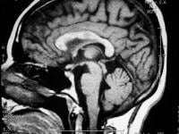 Sagittal
T1-weighted MRI depicts multiple hypointense lesions in the corpus
callosum; this finding is characteristic of multiple sclerosis.
Sagittal
T1-weighted MRI depicts multiple hypointense lesions in the corpus
callosum; this finding is characteristic of multiple sclerosis. 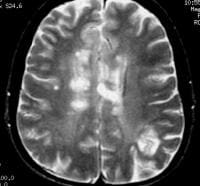 Axial
T2-weighted MRI in a patient with multiple sclerosis demonstrates
numerous white matter plaques in a callosal and pericallosal white
matter distribution.
Axial
T2-weighted MRI in a patient with multiple sclerosis demonstrates
numerous white matter plaques in a callosal and pericallosal white
matter distribution. 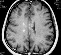 Axial
T1-weighted, gadolinium-enhanced MRI in a patient with multiple
sclerosis demonstrates several intensely enhancing pericallosal white
matter lesions compatible with active disease.
Axial
T1-weighted, gadolinium-enhanced MRI in a patient with multiple
sclerosis demonstrates several intensely enhancing pericallosal white
matter lesions compatible with active disease. 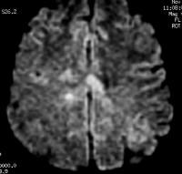 Axial
diffusion-weighted MRI in a patient with multiple sclerosis shows
several hyperintense lesions, a feature of inflammatory disease
activity.
Axial
diffusion-weighted MRI in a patient with multiple sclerosis shows
several hyperintense lesions, a feature of inflammatory disease
activity. 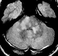 Axial
proton density–weighted MRI through the posterior fossa in a patient
with multiple sclerosis demonstrates multiple bright foci in the
brainstem and cerebellum. Proton density–weighted sequences are highly
sensitive for the detection of plaques in multiple sclerosis, especially
in the posterior fossa.
Axial
proton density–weighted MRI through the posterior fossa in a patient
with multiple sclerosis demonstrates multiple bright foci in the
brainstem and cerebellum. Proton density–weighted sequences are highly
sensitive for the detection of plaques in multiple sclerosis, especially
in the posterior fossa. 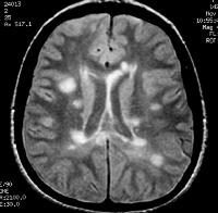 Axial
proton density–weighted MRI demonstrates multiple lesions in a
distribution characteristic of multiple sclerosis. Specifically, the
periventricular lesions and the more peripheral white matter lesions
near the gray matter–white matter junction are typical MRI findings in
multiple sclerosis.
Axial
proton density–weighted MRI demonstrates multiple lesions in a
distribution characteristic of multiple sclerosis. Specifically, the
periventricular lesions and the more peripheral white matter lesions
near the gray matter–white matter junction are typical MRI findings in
multiple sclerosis. 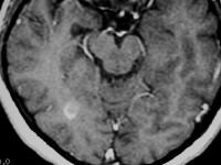 Axial
T1-weighted, gadolinium-enhanced MRI in a patient with multiple
sclerosis depicts enhancement of a plaque in the right temporo-occipital
lobe, signifying disease activity. Note the C-shaped, or arclike,
enhancement, which is fairly characteristic of multiple sclerosis.
Axial
T1-weighted, gadolinium-enhanced MRI in a patient with multiple
sclerosis depicts enhancement of a plaque in the right temporo-occipital
lobe, signifying disease activity. Note the C-shaped, or arclike,
enhancement, which is fairly characteristic of multiple sclerosis. 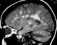 Sagittal
proton density–weighted MRI in a patient with multiple sclerosis
demonstrates the characteristic corpus callosal and pericallosal white
matter lesions.
Sagittal
proton density–weighted MRI in a patient with multiple sclerosis
demonstrates the characteristic corpus callosal and pericallosal white
matter lesions. 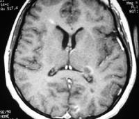 Axial
T1-weighted, gadolinium-enhanced MRI in a patient with multiple
sclerosis depicts several enhancing lesions, at least 2 of which show
characteristic C-shaped, or arclike, peripheral enhancement.
Axial
T1-weighted, gadolinium-enhanced MRI in a patient with multiple
sclerosis depicts several enhancing lesions, at least 2 of which show
characteristic C-shaped, or arclike, peripheral enhancement.  Axial
diffusion-weighted MRI in a patient with multiple sclerosis shows
several hyperintense lesions, a feature of inflammatory disease
activity.
Axial
diffusion-weighted MRI in a patient with multiple sclerosis shows
several hyperintense lesions, a feature of inflammatory disease
activity. Proton density (PD)–weighted MRI has an advantage
over standard T2 imaging, because on PD series, MS lesions remain
hyperintense, while the CSF signal is suppressed. Therefore, the lesions
are easily identified. Depending on the PD technique, the CSF signal is
suppressed to a variable degree, rendering it isointense to hypointense
relative to the brain parenchyma. This sequence results in substantial
suppression of Virchow-Robin spaces, which are perivascular CSF spaces
that may penetrate to the subcortical white matter. These spaces may
appear as hyperintense spots on standard T2-weighted MRI scans.
Compared
with other techniques, nonenhanced T1-weighted MRI is far less
sensitive in detecting MS lesions. Acute lesions usually are not
depicted at all. With T1-weighted MRI, the clinician can gain a general
appreciation of the global cerebral atrophy that occurs with advanced
chronic MS. Global atrophy has been suggested to have the strongest
imaging correlation with disability.
Chronic MS lesions usually
result in localized leukomalacia, and they may appear as hypointense
lesions that represent loss of tissue.
Gadolinium-enhanced
T1-weighted MRI scans can depict acute, active MS lesions. These appear
as enhancing white matter lesions; the presence of an enhancing lesion
has been shown to increase the specificity for MS.
[2, 18]
FLAIR MRI
Newer
MRI pulse sequences and techniques, including fluid-attenuated
inversion recovery (FLAIR) MRI and MR spectroscopy, have emerged that
are potentially useful in the evaluation of patients with MS.
FLAIR
MRI is a heavily T2-weighted technique that dampens the ventricular
(ie, free-water) CSF signal. Thus, the highest signals on the sequence
are from certain brain parenchymal abnormalities, such as MS lesions,
while the CSF appears black. This appearance is different from that on
PD-weighted MRIs, on which periventricular MS lesions may appear nearly
isointense to the adjacent CSF. (See the image below.)
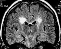 Coronal
fluid-attenuated inversion recovery (FLAIR) MRI in a patient with
multiple sclerosis demonstrates periventricular high–signal intensity
lesions, which exhibit a typical distribution for multiple sclerosis.
FLAIR MRI is a highly sensitive sequence for lesion detection,
particularly supratentorially.
Coronal
fluid-attenuated inversion recovery (FLAIR) MRI in a patient with
multiple sclerosis demonstrates periventricular high–signal intensity
lesions, which exhibit a typical distribution for multiple sclerosis.
FLAIR MRI is a highly sensitive sequence for lesion detection,
particularly supratentorially. The greater relative
suppression of CSF on FLAIR images compared with PD-weighted series
increases the contrast between periventricular lesions and CSF,
enhancing their detection. FLAIR has been shown to be superior to
PD-weighted sequences in the detection of MS lesions in the cerebral
hemispheres. However, PD-weighted imaging remains the investigation of
choice for infratentorial lesions.
[28]
MR spectroscopy
Magnetic
resonance (MR) spectroscopy uses the characteristic spectra of specific
biochemical markers to quantitate organic compounds in vivo.
N
-acetylaspartate (NAA) is a relatively specific neuronal marker that is
present in sufficient concentrations in the brain to be revealed on MR
spectroscopic images. By comparing the spectral signal of NAA with that
of creatinine (Cr), MR spectroscopic can be useful in assessing neuronal
and axonal loss.
Arnold et al noted that the NAA-Cr ratio in the
CNS was decreased in moderate to advanced MS. White matter that
appeared normal on T1- and T2-weighted images also demonstrated the
reduction.
[29] In
addition, a normal ratio was noted in the area of a recently active
lesion associated with clinical deficits that subsequently resolved. The
findings led the authors to propose that MR spectroscopic findings may
be able to help identify irreversible axonal damage.
In a study
involving 88 patients with MS, De Stefano et al found a strong
correlation between disability scores and NAA-Cr ratios.
[30] The
ratio exhibited a stronger correlation in patients with MS patients who
had milder disability scores. Because MR spectroscopy appears to be
capable of depicting changes in white matter that are not detected with
routine pulse sequences and because the findings are correlated with
disability scores, the use of MR spectroscopy may prove valuable in
monitoring patients after treatment and in formulating their prognosis.
Nonstandard MRI sequences
Beyond
the standard MRI sequences that are used in clinical practice (T1 +/-
Gad, T2, diffusion-weighted imaging, FLAIR), more advanced MRI
techniques have been used for research purposes for several years. Many
of these series require greater magnetic field strengths over the
popular 1.5T, but with the increasing availability of 3T MRI, these
sequences will likely find their way more and more into standard
clinical practice.
Diffusion tensor imaging (DTI) can utilize
diffusion-weighted imaging techniques in different orientations to
establish pathology along white matter tracts in the CNS. DTI can
identify demyelination and loss of axons along tracts that would
otherwise go undetected by conventional techniques.
[31, 32, 33] DTI
can also identify disease activity in and injury to gray matter
structures, which in turn can be used as a markers of disease activity
and severity.
[34, 35, 36, 37]
Double
inversion recover (DIR) sequences can also detect cortical lesions with
increased sensitivity over standard MRI sequences, with higher MRI
field strengths improving sensitivity.
[38]
Magnetization
transfer imaging (MTI) is capable of identifying MS lesions before they
can be detected by conventional MRI techniques.
[39, 40]
Limitations
In
virtually all patients with clinically well-established MS, MRI scans
demonstrate the corresponding changes. False-negative findings occur
more frequently in patients with early MS and a minimal clinical history
of neurologic impairment than in other patients.
O'Riordan et al prospectively found that in 3 of 27 patients with normal MRI findings, MS subsequently developed.
[17] However,
the patients with normal MRI findings all developed lesions detectable
on MRI scans when the disease became established. Similarly, as patients
are followed for longer periods, the rate of false-positive findings
decreases, because in many patients with abnormal MRI findings after a
single neurologic event, the clinical criteria for MS eventually
develop.
Gadolinium-based contrast agents have been linked to the
development of nephrogenic systemic fibrosis (NSF), also called
nephrogenic fibrosing dermopathy (NFD). The disease has occurred in
patients with moderate to end-stage renal disease after being given a
gadolinium-based contrast agent to enhance MRI or MR angiography scans.
NSF/NFD is a debilitating and sometimes fatal disease. Characteristics
include red or dark patches on the skin; burning, itching, swelling,
hardening, and tightening of the skin; yellow spots on the whites of the
eyes; joint stiffness with trouble moving or straightening the arms,
hands, legs, or feet; pain deep in the hip bones or ribs; and muscle
weakness.